Nikon Confocal Microscope Facility
Biomedical Sciences Resources
Jeff Thuma, Manager
thuma@ohio.edu
General Description
The Heritage College Microscopy Core Facility houses two microscopes (Nikon E600 conventional widefield and Nikon A1R confocal) and a dedicated offline workstation for image analysis. These powerful systems permit capture of high-quality images of fixed or living tissues, cells and molecules. A sample preparation area is available adjacent to the microscope room. This facility can accommodate a variety of specimens and assist a broad range of users.
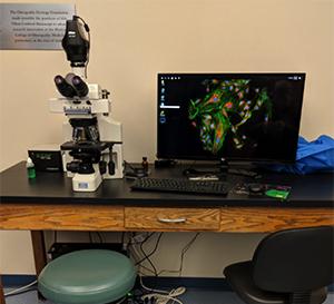
Nikon E600 Widefield Microscope
- Nikon E600 upright microscope
- LED illumination for both transmitted and reflected (fluorescence) applications
- Six objective lenses
- 10x Plan phase contrast (Air)
- 20x Plan Apo DIC (Air)
- 40x Plan Apo DIC (Air)
- 40x Plan Fluor DIC (Air), long working distance
- 60x Plan Apo DIC (Oil)
- 100x Plan Apo DIC (Oil)
- Three filter sets (blue, green, red)
- 16.25 Megapixel monochrome camera
- NIS Elements software, which integrates data acquisition and image analysis
Fees
- $5 per hour (Internal; Operated by user)
- $10 per hour (External; Operated by user)
Nikon A1R Confocal Microscope
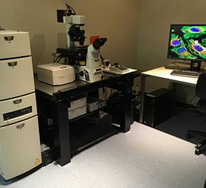
- Nikon Eclipse Ti inverted microscope
- Motorized XY stage
- Nikon's patented PFS Perfect Focus System to compensate for focal drift during time lapse and extended experiments
- High-speed piezo Z stage stepper for rapid Z-stack acquisition (100 um/s)
- LED illumination for both transmitted and reflected (fluorescent) light applications
- Five objective lenses
- 10x Plan Apo (Air)
- 20x Plan Apo DIC (Air)
- 40x Plan Fluor DIC (Oil)
- 60x Plan Apo Lambda DIC (Oil)
- 100x Plan Apochromat Lambda DIC (Oil)
- Four lasers: 405nm, 488nm, 561nm, 640nm
- Two types of scanners: galvanometric and resonant
- The galvo scanner scans at ~1 frame per second at 512 x 512 pixels with a maximum image size of 4096 x 4096 pixels
- The resonant scanner can scan up to 30 frames per second at 512 x 512 pixels
- DU4 detector with four detection channels in the 400-820nm range; channels 2 and 3 are Gallium-Arsenide-Phosphide (GaAsP) detectors, which are very sensitive in the 500-600nm range and improve signal-to-noise ratio
- 32-channel spectral detector with wavelength resolution at 10nm/6nm/2.5nm gratings, allowing for separation of closely overlapping spectra and elimination of autofluorescence
- Stage-mounted environmental chamber is available to control temperature, humidity and gas during live imaging experiments
- NIS Elements software, which integrates data acquisition and image analysis
- Additional software upgrades including 3D deconvolution, calcium & FRET, and object tracking
Fees
- $25 per hour (Internal; Operated by user)
- $60 per hour (External; Operated by user)
Details on Using Facility
Services Provided
Training in proper use and care of the microscope is mandatory for all users and will be conducted by facility staff. Basic training usually can be completed in 1-2 hours. Additional training is required for more advanced techniques (including the live cell incubation chamber). Users are expected to prepare and analyze their own samples independently; however, further assistance is available upon request.
Application
To request an application for a training session, please contact Jeff Thuma, thuma@ohio.edu.
Location
Academic and Research Center, Room 246
Access and Operating Guidelines
The facility is available to the entire Ohio University research community as well as external users. All users must begin by completing an application form. The application form will be reviewed by the director and, upon approval, the user will be contacted by the manager to schedule one-on-one training using their specimens. All users must complete training with the manager before being authorized to use the equipment.
The equipment is available by appointment only using a reservation website. Access to equipment and website are granted upon completion of training.
User Responsibilities
It is the user's responsibility to prepare samples. Each user is also responsible for supplying details of any hazards (e.g., infectious organisms, blood-borne pathogens, toxic chemicals) associated with the preparation in accordance to Ohio University Laboratory & Radiation Safety regulations (740.593.1666). All users are required to read the Care and Use of Objective Lenses document for the microscope.
Publication and Acknowledgements - Very Important
To accurately reflect the assistance this resource played in your research and to help ensure its continued funding, please include the following statement in all publications: "We acknowledge use of the Ohio University Heritage College of Osteopathic Medicine Microscopy Core."
Acknowledgement and Co-authorship Policies
Co-authorship on publications for staff of the Microscopy Core may be considered appropriate when they have provided one or more of the following:
- Significant intellectual contribution to the design of the published experiments
- Substantial practical contribution to the generation, analysis and/or interpretation of experimental data
- Indispensable technical support that contributed intellectually or scientifically to the advancement of the work
Staff who have made an intellectual or technical contribution not justifying authorship as defined above should be given appropriate acknowledgement in any resulting publications. This provides evidence to demonstrate our involvement in the research of multiple laboratories, which will help us retain dedicated support staff, maintain the equipment and procure new equipment in the future.
Problem Management
Manager
Jeff Thuma
740.593.0680 (lab)
014 Wilson Hall West
thuma@ohio.edu