
903 Peripheral IV Catheter Placement and Management
| Document Name: Peripheral IV Catheter Placement and Management | Document Number: 903 | Revision Date: 13AUG2024 | Supersedes: 605.00 |
| Target Group: Research Personnel | Approval Date: 20Dec2024 | Effective Date: 01Jan2025 | Training Required: -Ohio credential to perform IV’s or place IV catheter. -Bloodborne Pathogens -Human Subjects Protection (HSP) Training -Good Clinical Practices (GCP) Training -Clinical Competency Training |
Purpose
To ensure the Principal Investigator (PI) and all research team members assisting with the conduct of clinical research at the Ohio University Heritage College of Osteopathic Medicine’s Clinical and Translational Research Unit (CTRU) are informed about their obligations and responsibilities as they pertain to Good Clinical Practices (GCP), the investigational plan, applicable regulations, and institutional policies.
This Standard Operating Procedure (SOP) describes the standards for the initiation and management of peripheral intravenous (IV) access with consideration of participant safety, comfort, and protocol needs. The initiation and management of peripheral intravenous (IV) access for serial blood draws and/or medication administration will be based upon protocol requirements.
Responsibilities
The CTRU is responsible for developing, implementing, and maintaining SOPs. Creating new or revising an existing SOP is determined by changes to federal regulation, guidelines, institutional policies, or procedures. All applicable documents will be provided to departments and research teams conducting human subject research.
Definitions
Peripheral IV access: IV access obtained through the superficial veins of the upper extremity.
Special Peripheral IV Access: With approval from the CTRU Medical Director the superficial veins of the lower extremity, or scalp veins of infants, are also acceptable locations for peripheral access. The lower extremity and scalp present additional risks and these warrant review by the CTRU Medical Director.
Infiltration: Accumulation of fluid in the tissue surrounding an IV needle site.
Phlebitis: Inflammation of the wall of the vein.
Air embolism: The obstruction of a blood vessel (usually occurring in the lungs or heart) by air carried via the bloodstream.
Hematoma: Swelling due to ruptured blood vessels, bruising will occur.
Procedures
1. Training
- In Ohio, under proper supervision, personnel can perform peripheral IVs as allowed by the state practice act for healthcare providers and/or their collaborative agreement with a supervising physician.
- Individuals performing peripheral IV must have completed IV training either on the job or through a course.
- Clinical competency in peripheral IV techniques requires individuals to perform at least 3 successful peripheral IVs under supervision before performing this procedure unsupervised.
- Staff must complete the Ohio University (OHIO) Bloodborne Pathogens Training before attempting peripheral IVs and annual refresher training as required by the OU Environmental Health and Safety Office.
2. Peripheral IV Considerations
- The needle/catheter gauge is determined by the credentialed staff based on protocol requirements, condition of veins, and number/amount of blood drawn, using the largest gauge feasible.
- Registered Nurses (RNs) may restart a peripheral line without a physician’s order for site changes due to infiltration, phlebitis, or the inability to get protocol-required blood draws from the site. Areas with open wounds or signs of infection should be avoided.
- Other credentialed staff may restart of peripheral line without a physician’s order if it is stated in their collaborative agreement with their supervising physician.
- An IV will not be attempted if the credentialed staff cannot locate an appropriate site, and/or if the participant expresses the desire to exit from the procedure.
- Ask if the subject has had problems with blood draws, allergies or sensitivities to adhesives, or a history of complications.
- IV catheters shall only be placed in the upper extremities.
- IV catheters should not be placed on the arm on the side on which a mastectomy has been performed or where there has been extensive injury/surgery.
- Avoid scar tissue, areas with a hematoma, areas of infection and areas with extensive burns.
- Once inserted, the IV fluids may be ordered for ‘keep vein open’ (KVO) with IV fluid or IV saline-lock depending on study protocol.
- The intravenous catheter will be removed before the participant is discharged from the Clinical & Translational Research Unit (CTRU).
- Consider recruiting assistance from a second staff member for assistance prior to start.
- Clean any extra equipment (i.e. heating pad, vascular visualization device, etc.) used during the procedure per manufacturer’s cleaning instructions.
3. Supplies Needed
- Gloves
- Angiocath
- Tagaderm® or non-adhesive dressing
- Gauze pad
- Antiseptic agent
- Single use tourniquet wrap (optional)
- If using a saline lock: 10cc of preservative-free normal saline, IV cap
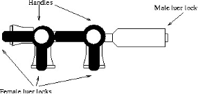
- If using stopcocks: IV tubing, IV solution, two stopcocks, extra gauze, IV pole
4. Peripheral IV Procedure
- Inspect the integrity of equipment.
- Assess the participant’s understanding of the study and the procedure(s) to be done at this visit.
- Explain procedure to participant.
- Wash hands thoroughly.
- If using stopcocks with IV drip: hook IV tubing set to fluid bag and prime tubing. Insert and prime stopcocks, if necessary, for the procedure.
- Select site considering the purpose and duration of the procedure.
- If palpation and/or visualization is difficult, consider the following actions:
- Consult with another credentialed staff, if available.
- Use a vascular visualization device, if available.
- Use a heating pad to warm the extremity if not contraindicated. (Contraindications include patients with peripheral vascular disease, bleeding disorder, local malignancy, acute inflammation or trauma, edema, infection, open wounds, over large scars, patients with impaired sensation or impaired ability to communicate.)
- Have the subject drink water to increase hydration if not contraindicated (i.e. fasting).
Apply tourniquet above intended IV site. Tie tourniquet in a manner to allow release with one hand. Remove the tourniquet ASAP--not to exceed 1 minute. Apply tourniquet above intended IV site. Tie tourniquet in a manner to allow release with one hand. Remove the tourniquet ASAP--not to exceed 1 minute. - Prepare insertion site by cleansing with Chloraprep® or alternative antiseptic (i.e. alcohol prep pad) using aseptic technique. Cleanse site with friction using Chloraprep® or alternative antiseptic and allow it to dry.
- Do not touch the prepped area with your fingers. If palpable reassessment of the vein needs to be done, re-prep the skin prior to venipuncture.
- Put on clean gloves.
- Anchor the skin 1-2 inches below the insertion site and insert angiocath at the prepared site.
- Look for blood return in the flashback chamber, advance the catheter, and release tourniquet while applying pressure to vein just above insertion site to prevent bleeding.
- If a credentialed staff is unable to start an IV after a thorough participant assessment and/or two attempts at venipuncture, consider the following actions:
- Another credentialed staff should be requested to assess and attempt IV.
- The study physician or PI (if physician) may attempt to start the IV with the verbal permission of the participant.
- If still unsuccessful or lacking the permission of the participant, the Principal Investigator (PI) will be notified of the difficulty in initiating a peripheral catheter and the study procedure will be aborted.
Saline Lock versus Stopcock Method
| Saline Lock | Stopcock Method | |
|---|---|---|
| 16. | Secure angiocath to skin using Tegaderm. | Attach extension tubing and IV tubing, if using, and open roller clamp. |
| 17. | Attach cap and flush with normal saline syringe then slide clamp while flushing. | Secure stopcocks to skin of forearm supporting them with folded sterile 2x2 gauze dressings and tape.
|
| 18. | Set drip rate per protocol |
- Assess site. Fluids should flow freely and evenly. Inspect/palpate around angiocath for swelling/rigidity as those are signs of infiltration.
- If infiltrated, immediately turn off fluids and discontinue IV.
- Document size of angiocath, location, date, and time in participant’s source document.
5. Discontinuing IV
- Participants with venous access must remain at the CTRU while the catheter is in place unless accompanied by a member of the study team.
- Discontinue IV per protocol, complications, or per participant’s request.
- Put on clean gloves.
- Clamp fluids/IV.
- Remove any tape.
- Gently remove the IV catheter.
- Visually inspect catheter to make sure tip is intact. If not, notify the study physician/PI.
- Place gauze over removal site and hold pressure for a few minutes.
- Monitor site for ongoing bleeding before placing bandage.
- Apply bandage or tape and cotton ball if an allergy exists.
6. General Guidelines for Flushing and Capping
- Prepare clean work area.
- Wash hands and put on gloves.
- Take the cap off the saline syringe. Do not touch the inside of the cap or the syringe tip to any surface.
- Hold the syringe so the open tip points to the ceiling. Gently tap the side of the syringe to move any air bubbles to the top.
- Push in the plunger until a drop of liquid appears at the open tip of the syringe.
- Use a new alcohol pad to clean the cap at the end of the IV line for 15-30 seconds. Allow to air dry.
- Open the clamp on IV line or extension set.
- Attach, push, and twist the end of the saline syringe onto the cap of your IV line.
- Push the plunger to flush the IV line using a start/stop action. Untwist the syringe to remove.
- Administer medication or obtain sample per protocol.
- Repeat the above steps for the saline flush.
- Remove the syringe and clamp the tubing.
- Attach cap per injection/needless system manufacturer guidelines.
7. Potential Complications
- Infiltration
- Caused by penetration of the vein wall by the needle/catheter or later dislodgment.
- Signs and Symptoms:
- The flow rate may or may not be slow or there is no flow of solution.
- Infusion site is cool and hard to the touch.
- Infusion site or extremity is pale and/or swollen.
- Subject complaints of pain, tenderness, burning, or irritation at infusion site.
- Fluid leaking around infusion site.
- Corrective Action:
- Stop IV infusion immediately and remove needle or catheter.
- Elevate extremity in which IV had been placed.
- If noticed within 30 minutes of onset, apply ice to swelling or per protocol.
- If noted later than 30 minutes of onset, apply warm compresses to encourage absorption or per protocol.
- Notify study physician/CTRU Medical Director of infiltration.
- Document observations and actions.
- Restart IV in another location, if directed.
- Preventative Measures:
- Tape catheter hub and tubing securely to limb.
- Stabilize extremity in use by applying arm board if necessary.
- Phlebitis
- Caused by the following:
- Injury to the vein during puncture.
- Needle movement after insertion.
- Irritation to vein as result of:
- Long-term therapy (vein overuse).
- Irritating or incompatible additive.
- Using large-bore catheters.
- Infection.
- Signs and Symptoms:
- Sluggish flow rate.
- Swelling around the infusion site.
- Patient complaints of pain and tenderness.
- Redness and warmth along the vein.
- Corrective Action:
- Stop IV infusion immediately.
- Report observations to study physician or CTRU Medical Director.
- Treat affected limb like an infiltration.
- Document observations/actions.
- Preventative Measures:
- Keep infusion flowing at prescribed rate.
- Select large vein when irritating drugs/fluids are given.
- Change IV site PRN.
- Instruct participants to continue to monitor IV insertion site for signs/symptoms of phlebitis once discharged from CTRU.
- Caused by the following:
- Air embolism
- Caused by the following:
- Failure to remove air from the tubing.
- Allowing the solution to run dry.
- Disconnected IV tubing.
- Signs and Symptoms:
- Abrupt drop in blood pressure.
- Weak, rapid pulse.
- Cyanosis.
- Chest pain.
- Sudden death.
- Corrective Action:
- Call 911.
- Notify PI and study physician/Medical Director immediately.
- Immediately place patient on left side with feet elevated to allow the pulmonary artery to absorb small air bubbles.
- Administer oxygen as directed by physician.
- Preventative Measures:
- Clear all air from tubing before attaching to the patient.
- Monitor solution levels closely and change before they are empty.
- Check to see that all connections are secure.
- Caused by the following:
- Infection
- Caused by the following:
- Use of contaminated equipment.
- Poor aseptic venipuncture technique.
- Contaminated site or IV equipment not changed regularly.
- Signs and Symptoms:
- Redness, swelling, and soreness around IV site.
- Sudden rise in temperature and pulse.
- Drainage from IV site.
- Corrective Action:
- Notify PI and study physician.
- Discontinue IV.
- Use strict aseptic technique when cleaning and dressing the wound.
- Document all corrective actions taken.
- Preventative Measures:
- Use complete aseptic technique when starting an IV.
- Clean site thoroughly when IV is initiated.
- Anchor catheter and tubing securely.
- Caused by the following:
- Hematoma
- Caused by the rupture of blood vessels.
- Signs and Symptoms:
- Bruising.
- Swelling.
- Pain.
- Corrective Actions:
- Stop procedure.
- Immediately remove tourniquet and IV.
- Apply pressure to venipuncture site until bleeding has stopped.
- Cold packs can be applied to reduce bruising and pain.
- Notify PI and study physician.
- Thrombophlebitis
- Caused by irritation of the vein leading the body to trigger clotting mechanisms.
- Signs and Symptoms:
- Red, swollen, irritated skin.
- Pain, warm, tender or hard sensations in the area (cord).
- Hardening of the vein.
- Fever.
- Corrective Actions:
- Discontinue IV.
- Elevate the extremity and apply a warm compress.
- Notify study physician.
- Preventative Measures:
- Avoid placing cannulas in places of flexion when possible.
- Use of pumps/controllers.
- Avoid cannulation in lower extremities.
8. Suggested Informed Consent Language
Definition: intravenous (IV)-within a vein.
IV Catheter-a small, flexible tube inserted in the vein
(choose A)
An intravenous small plastic catheter or tube (IV) will be placed in your arm for the collection of blood samples. The IV line will make it easier to collect the necessary number of blood samples.
<insert how much blood and when it will be collected>. Most of this blood will be used to measure <specify what>.
STUDY PROCEDURE RISKS
During the collection of blood samples, you may experience pain and/or bruising at the IV site. Although rare, localized clot formation, air embolism and infections may occur. Lightheadedness and/or fainting may also occur during or shortly after IV placement. There is the potential to have more than one attempt to insert the IV.
(choose B)
To make taking blood easier, and prevent multiple ‘sticks’, a catheter (small diameter plastic tube about 1 inch long) will be placed into a vein in your arm. The total amount of blood taken during this test will not be more than <insert how much blood and when it will be collected>. Although uncommon, it is possible that the initial arm catheter will stop working properly during the procedure (we will no longer be able to take blood samples from it). If this occurs, it will be removed, and we will ask you if we have permission to place a second catheter in a different part of your arm or perform single needle stick(s) so that we can continue taking blood samples.
References: None
Attachments: None
Appendices: None
Approver: Director, CTRU and Clinical Partnerships